Case Study: FNA of pancreatic cystic lesion

Introduction

High equipment costs have confined EUS mainly to hospital settings, limiting access.  The EndoSound Vision System™ (EVS™) technology transforms any endoscope into a fully functioning endoscopic ultrasound (EUS) system.  We use the EVS for Echo-EGD, defined as diagnostic EGD enhanced with ultrasound for many imaging-only procedures in our ASC.  This paper describes the EVS’s additional capabilities of fine needle aspiration (FNA) which is traditionally performed in a hospital setting.

The EVS is cost-effective and opens new possibilities for EUS across care settings.  EndoSound provides needles that fit the length of gastroscopes enabling the capability of FNA in the ASC.  Thus, given the cost of the traditional EUS equipment this is an exciting innovation to perform EUS procedures in smaller surgery centers.

Below, is a case using the EVS to evaluate the symptom and perform FNA of a pancreatic cyst.

Patient History

81-year-old male with history of lymphoplasmacytic lymphoma and pancreatic surgery for pancreatic cyst many years ago presented with upper abdominal pain. Details of prior surgery is not known.

The Procedure

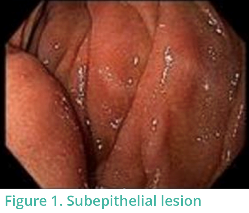
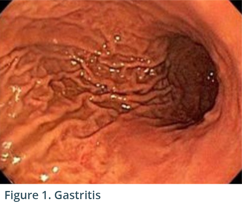
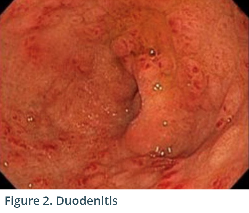
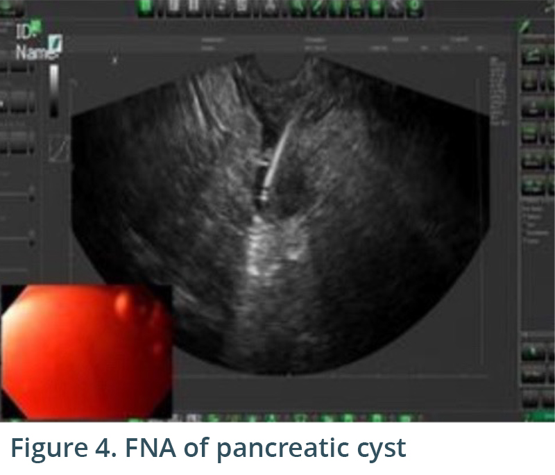
Endoscopic exam revealed gastritis (Fig 1) and duodenitis in duodenal bulb (Fig 2). Pancreatic parenchyma was without mass, and there was no dilation of main pancreatic duct.  There was a cyst in head of pancreas measuring 4.7 x 2.5 mm and another in the body of pancreas measuring 5.1 x 4.3 mm. Both cysts were without mural nodule or associated mass and were too small for meaningful FNA. Tail of pancreas had a 14.6 x 12.9 mm cyst without mural nodule or mass status post FNA with 22G needle (Figs 3, 4) with aspiration of approximately 1.5-2 cc of viscous/straw colored fluid sent for analysis. Other etiologies of abdominal pain such as gallstones and biliary causes were ruled out during EUS (Figs 5,6).

Cytology was negative for malignancy. CEA was 3008 and amylase 105.  Molecular test was negative for GNAS, KRAS and LOH and concluded that there was a 97% probability that the cyst would remain benign over the next three years.  Duodenal biopsy revealed gastric heterotopia, and gastric biopsy was negative for helicobacter pylori.  It was determined that the most likely etiology of his abdominal pain was gastric heterotopia versus Zanubrutinib which he was taking for his lymphoma.

Conclusion

The EVS allowed both visualization/fine needle aspiration of pancreatic cyst lesion and the work-up of patient’s abdominal pain in an ASC setting.  With the supply of the FNA needles for the EVS specifically, it is a complete system allowing FNA or fine needle biopsy (FNB) in the ASC.  Moreover, Echo-EGD can be utilized as a first-line investigation, bypassing other imaging modalities and streamlining the diagnostic process (Sahai, et al., 2000; Chang et al., 2010).  By making EUS capabilities more accessible and affordable, we are revolutionizing the standard of care in endoscopy.


References:

Sahai et al., Gastrointest Endosc 2000;52:153-9. EUS to detect evidence of pancreatic disease in patients with persistent or nonspecific dyspepsia.

Chang et al., Gastrointest Endosc 2010; 72: 967-74. EUS compared with endoscopy plus transabdominal US in the initial diagnostic evaluation of patients with upper abdominal pain.

Case Study: Windsock diverticulum

Introduction

Echo-EGD, which I define as diagnostic EGD enhanced with ultrasound, is perhaps ideal to maximize the find rate.  Today, it is more accessible than ever and with it comes the promise to improve patient care and reduce healthcare spending.

We believe that Echo-EGD is more possible and necessary than ever because:

  • There are 8 million EGDs performed annually in the U.S.
  • A significant portion could benefit from a more comprehensive exam compared to standard EGD.
  • It can often reduce the need for additional cross-sectional studies such as CT or MR.
  • Echo-EGD can be performed in the ASC.

High equipment costs have confined EUS mainly to hospital settings, limiting access.  The EndoSound Vision System™ (EVS™) technology transforms any endoscope into a fully functioning EUS system. EVS is cost-effective and opens new possibilities for Echo-EGD across care settings.

Below I describe a case where Echo-EGD with EVS helped achieve a definitive diagnosis.

Patient History

A 50-year-old male with reflux and dysphagia was found to have a subepithelial lesion in the second part of the duodenum during EGD.  The patient was referred for EUS for further investigation.

The Procedure

A subepithelial lesion was identified in the second portion of duodenum (Figure 1). Proximal to this lesion, there was a diverticulum with a large opening (Figure 2). To have an optimal angle for endosonography based on the endoscopic location of the lesion, the ultrasound probe was placed at 7 o’clock position (Figure 3). EUS confirmed that the “subepithelial lesion” seen on EGD was the outer portion of a windsock-like diverticulum. Water infusion in duodenal lumen resulted in water flowing into the inner part of diverticulum on EUS (Figures 4, 5). Figure 5 shows the sonographic image of a windsock diverticulum as seen on EUS with the picture of an actual windsock inserted for comparison.

Conclusion

Confirmation of a windsock diverticulum masquerading as a subepithelial lesion was achieved by performing EUS with EVS. Even though this case was a referral from another gastroenterologist, it could have easily been an Echo-EGD case should the lesion have been found by an endosonographer with access to EVS at the time of initial EGD. This, in turn, would have enabled the delivery of efficient care cost-effectively. Another advantage of EVS compared to traditional EUS scopes is that the probe placement can be tailored to the location of the lesion viewed endoscopically. In our case, the lesion was in 7 o’clock position and thus the probe was placed at 7 o’clock position accordingly.  Lastly, the forward viewing scope orientation reduced the risk of accidentally entering the diverticulum and causing perforation – a complication occasionally seen with side-viewing endoscopes.

This shift in approach may lead to changes in our patient flow. Specifically, we may now opt for Echo-EGD as a first-line investigation, bypassing other imaging modalities and streamlining the diagnostic process (Sahai et al., 2000).  More recently, additional data supports this approach that Echo-EGD is a viable option for patient care (Chang et al., 2010).  By making EUS capabilities more accessible and affordable, we are not just introducing new technology – we are revolutionizing the standard of care in endoscopy.


References:

Sahai et al., Gastrointest Endosc 2000;52:153-9. EUS to detect evidence of pancreatic disease in patients with persistent or nonspecific dyspepsia.

Chang et al., Gastrointest Endosc 2010; 72: 967-74. EUS compared with endoscopy plus transabdominal US in the initial diagnostic evaluation of patients with upper abdominal pain.